Tailor’s Bunion

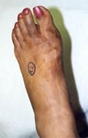
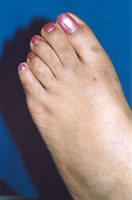
A Tailor's Bunion (Bunionette) is a type of bunion that occurs on the outside of the foot at the base of the little toe. It is a prominence that may be caused by irritation of bone or soft tissue about the lateral aspect of the fifth metatarsal head. Symptoms result from shoe pressure over the bony prominence, local nerve irritation, capsulitis, or a bursa.

Tailor's Bunion is named from clothing tailors sitting with crossed legs.

There is a three to one female to male predominance. They are common in adolescents and adults. Two thirds of patients have significant pes planus (flat feet).

As far as anatomical pathology is concerned, constricting footwear over the lateral 5th metatarsal head yields pain and a bursa. There may be incomplete insertion or development of the transverse metatarsal ligament.

Hyperactive Abductor digiti minimi and Opponens digiti minimi muscles may be present. One may encounter insufficient insertion of the Adductor Hallucis muscle. One may posess a congenital wide 5th metatarsal head.

On clinical presentation, there is pain and irritation at the lateral 5th metatarsal head and prominence. An inflamed bursa may be present. Hyperkeratosis and erythema may be present over the 5th metatarsal head. The 5th toe assumes a varus attitude.

Radiological findings include rotation of the lateral plantar tubercle into a lateral position, increased intermetatarsal angle (IM)(normal 6.47degrees)-People with a tailor's bunion have an IM of 8.71 or greater, increased lateral deviation angle (normal 2.64) people with a tailor's bunion have a lateral deviation of 8.05 degrees, a large dumbbell-shaped 5th metatarsal head, arthritic changes resulting in exostosis (spur) formation at the 5th metatarsophalangeal joint, and any combination of the above conditions.

Type 1 Tailor's Bunion has enlargement of the lateral portion of the 5th metatarsal head. It approximately occurs in 27% of the cases.

Type 2 Tailor's Bunion results in lateral bowing of the diaphysis (shaft) of the 5th metatarsal. This occurs in approximately in 23% of the cases.

Type 3 Tailor's Bunion results in an increase in the 4th-5th intermetarsal angle. This occurs in 50% of the cases. Patients are most symptomatic with this type. Type 4 results from a combination of two or more of the above deformities and is frequently seen in rheumatoid arthritis patients.

Conservative treatments include wearing wide toe box shoes, debridement of lesions. Orthotics may be utilized to control pressure areas and mechanics of the foot. Non Steroidal Anti-Inflammatory Drugs may be taken. Injections may be given.

Surgical Treatment is indicated when conservative treatment fails. It is primarily indicated in special demands like sports. The goal is to decrease the width of the foot and decrease the pain and prominence of the Bunionette.

References

  • Banks, Alan S. et al. McGlamry's Comprehensive Textbook of Foot and Ankle Surgery. Philadelphia: Lippincott Williams and Wilkins, 2001.
  • Fallat LM, Buckholz J. Analysis of the tailor's bunions by radiographic and anatomical display. J AM Podiatry Assoc 1980;70:597-603.

Athlete’s Foot

Overview

Athlete's foot is a skin disease caused by a fungus, usually occurring between the toes.

The fungus most commonly attacks the feet because shoes create a warm, dark, and humid environment which encourages fungus growth.

Athlete's foot (tinea pedis) is a common, persistent infection of the foot caused by a dermatophyte, a microscopic fungus that lives on dead tissue of the hair, toenails, and outer skin layers. These fungi thrive in warm, moist environments such as shoes, stockings, and the floors of public showers, locker rooms, and swimming pools. Athlete's foot is transmitted through contact with a cut or abrasion on the plantar surface (bottom) of the foot. In rare cases, the fungus is transmitted from infected animals to humans.

Dermatophyte (skin) infections cause raised, circular pimples or blisters that resemble the lesions caused by ringworm. The infections are named after the part of the body they infect. Tinea pedis, therefore, refers to an infection of the feet.

Incidence and Prevalence

Athlete's foot is most common in men from the teenage years to the early 50s. Prevalence is affected by personal hygiene and daily activity. People with compromised immune systems are at greater risk.

Causes

There are at least four dermatophytes that can cause tineas pedis. The most common is trichophyton rubrum.

Signs and Symptoms

There are four common forms of athlete's foot:

  1. The most common is an annoying, persistent itching of the skin on the sole of the foot or between the toes (often the fourth and fifth toes). As the infection progresses, the skin grows soft. The center of the infection is inflamed and sensitive to the touch. Gradually, the edges of the infected area become milky white and the skin begins to peel. There may also be a slight watery discharge.
  2. In the ulcerative type, the peeling skin becomes worse. Large cracks develop in the skin, making the patient susceptible to secondary bacterial infections. The infection can be transmitted to other parts of the body by scratching, or contamination of clothing or bedding.
  3. The third type of tinea infection is often called "moccasin foot." In this type, a red rash spreads across the lower portion of the foot in the pattern of a moccasin. The skin in this region gradually becomes dense, white, and scaly.
  4. The fourth form of tinea pedis is inflammatory or vesicular, in which a series of raised bumps or ridges develops under the skin on the bottom of the foot, typically in the region of the metatarsal heads. Itching is intense and there is less peeling of the skin.

People with acute tinea infections may develop similar outbreaks on their hands, typically on the palms. This trichophyde reaction, also known as tineas manuum, is an immune system response to fungal antigens (antibodies that fight the fungal infection).

Diagnosis

Diagnosis is made by visual observation of the symptoms. The podiatrist eliminates the possibility of a bacterial infection by performing a microscopic examination of skin scrapings to determine the type of fungus causing the infection. Other tests include growing a fungal culture from skin scrapings and examining the patient's foot under an ultraviolet light.

Treatment

Tinea infections may disappear spontaneously or persist for years. They are difficult to eliminate and often recur. Best results usually are obtained with early treatment before the fungal infection establishes itself firmly. Antifungal drugs may be used to fight the infection.

If the infection is bacterial, a course of oral antibiotics may be prescribed.

If you are experiencing any of the symptoms addressed, we strongly recommend that you seek the advice of your podiatrist for proper diagnosis.

Nail Fungus

Fungal infection of the toenails is also known as onychomycosis. It is a common health problem that many of its sufferers do not know they have because the infection can be present for many years without causing any pain.

Onychomycosis is an infection of the plate and soft nail bed under the toenail, and is caused by dirt, debris, and different types of fungi.

Fungus attacks the nail and thrives on the nail's protein matter, keratin. Once this occurs, the nail will thicken, become discolored, foul smelling, and may even detach from the nail bed or deteriorate.

If ignored, the infection can spread to other toenails, the fingernails, and the skin. Its spread could also impair your ability to walk.

Proper foot hygiene and regular inspection of the feet and toes are the best ways to prevent this condition.

You should also:

  • Keep your bath or shower area clean
  • Wear shower shoes whenever possible, in public showering facilities
  • Wash your feet with soap and water every day
  • Dry your feet well, particularly between the toes
  • Always wear clean socks
  • Change shoes after two days to give them time to dry out
  • Use a foot powder daily
  • Properly disinfect pedicure tools and do not apply polish to any nails suspected of infection
  • Nails should be trimmed short and straight across
  • Visit your podiatrist annually

Treatment may vary depending on the nature of your infection. Your podiatrist may devise a treatment plan that includes one or all of the following:

  • Debridement (removal of diseased nail matter)
  • Prescription of an oral or topical medication
  • In severe cases, removal of the diseased nail

For more information on this subject, or to schedule an appointment with a footDrHorsley Podiatrist, please call (877) 372-6048

If you are experiencing any of the symptoms addressed, we strongly recommend that you seek the advice of your podiatrist for proper diagnosis.

Runners Feet

Black toe is a black discoloration beneath one or more toenails. It is usually caused by trauma resulting in bleeding under the nail plate. Swelling usually occurs and the nail could fall off.

There are numerous causes for a bruised toe, or black toenail. The main reasons for this are: their feet are unstable in their shoes and they are pronating and elongating (when weight is placed on the feet they stretch longer). The second reason would be that the shoe is improperly sized and fitted and the foot is sliding forward in the shoe. The third reason is a toe box that does not match the shape of the toe region on the foot. If the toe box is too narrow in height and it actually puts a downward pressure on top of the toenails, then eventually, over a long period of time, this can bruise the toenail and also make it turn black.

It is very common with runners to see black toenails. Their feet slide forward to the front of the shoe and the ends of the toes hit the front of the toe box of the boot. This can push the toenail backwards and bruise the root area from which the nail grows (the matrix).

The toenail will progressively turn black until it falls off, and a new toenail will start to grow underneath the old bruised toenail. This can take up to three to six months. If bleeding and pressure continues to build beneath the nail it is recommended that you seek the help of your podiatrist.

For more information on this subject, or to schedule an appointment with a footDrHorsley Podiatrist, please call (877) 372-6048 (Toll Free)

If you are experiencing any of the symptoms addressed, we strongly recommend that you seek the advice of your podiatrist for proper diagnosis.

Common Causes, and Symptoms of Foot Pain

Cause Location of Symptoms Symptoms
Corns and calluses Around toes, usually little toe, bottom of feet or areas exposed to friction Hard, dead, yellowish skin
Ingrown toenails Toe nails Nail curling into skin causes pain, swelling, and, in extreme cases, infection
Bunions and bunionettes (tailors bunion) Big toe (bunions) or little toe (bunionettes) Toes point inward
Area next to bony bump is red, tender, occasionally filled with fluid
Toe joint may be inflamed
Note: Osteoarthritis may also occur in the big toe in older people
Morton's neuroma Third and fourth toes and bottom of foot near these toes Cramping and burning pain around the third and fourth toe
The neuroma may be detected by pressing top to bottom using one hand and with the other hand pressing on the top of the foot and moving it side to side
Aggravated by prolonged standing and relieved by the removal of the shoes and forefoot massage
Hammertoe Usually second toe but may develop in any or all of the three middle toes Toes form hammer or claw shape
No pain at first, increasing as tendon becomes tighter and toes stiffen
Metatarsal stress fracture Area beneath the second or third toe Sudden pain when injury occurs
Sesamoiditis Ball of foot beneath big toe Pain and swelling
Plantar fasciitis Back of the arch right in front of heel At onset, some people report a tearing or popping sound
Pain, most severe with first steps after getting out of bed, decreasing after stretching, returning after inactivity
Bursitis of the heel Center of the heel Pain, with warmth and swelling. Increases during the day
Haglund's deformity (Pump bump) Fleshy area on the back of the heel Tender swelling aggravated by shoes with stiff backs
Stress fracture or, uncommonly, heel spurs Bottom of heel Sharp stabbing pain
Tarsal tunnel syndrome Anywhere along the bottom of the foot Numbness, tingling, or burning sensations, pain, most commonly felt at night
Flat feet The arch No arch
Often no pain or discomfort
Sometimes people report fatigue, pain, or stiffness in the feet, legs, and lower back
High Arches (Hollow feet) The arch High arches
Lower back pain, possible tendency to lower limb injuries
Achilles tendinitis Achilles tendon: area along the back between calf muscles and heel Pain worsens during physical activities (particularly running) after which the tendon usually swells and stiffens
If it ruptures, popping sound may occur followed by acute pain similar to a blow at the back of the leg

Foot Health FAQ

The Foot

Feet

The foot is a complex structure of 26 bones and 33 joints layered with an intertwining web of 126 muscles, ligaments, and nerves. The average person spends four hours on their feet and takes between 8,000 and 10,000 steps each day. The feet are very small relative to the rest of the body, and the impact of every step exerts tremendous force upon them -- about 50% greater than the person's body weight. During an average day the feet support a combined force equivalent to several hundred tons. In addition to supporting weight, the foot acts as a shock absorber and as a lever to propel the leg forward, and it serves to balance and adjusts the body to uneven surfaces. It is not surprising, then, that about 75% of Americans experience foot pain at some point in their lives. According to a recent study, chronic and severe foot pain is a serious burden for one in seven older disabled women. To compound problems, the lower back is often affected by injuries or abnormalities in the feet.

What Is Foot Pain?

Foot pain is generally defined by one of three sites of origin: the toes; the front of the foot (forefoot); or back of the foot (hind foot). Toe problems most often occur because of the pressure imposed by ill-fitting shoes. Pain originating in the front of the foot usually involves the metatarsal bones (five long bones that extend from the front of the arch to the bones in the toe) and the sesamoid bones (two small bones imbedded at the top of the first metatarsal bone, which connects to the big toe). Pain originating in the back of the foot can affect parts of the foot extending from the heel, across the sole (known as the plantar) to the ball of the foot.

General Conditions Causing Foot Pain

The causes of most incidents of foot pain are poorly fitting shoes. High-heeled shoes are major culprits for aggravating, if not causing, problems in the toes, where the most pressure is exerted. Other conditions can also cause or exacerbate foot pain. Weather affects the feet; they contract in cold and expand in hot weather. Foot size can also increase by 5% over the day and change shape and size depending on whether a person is walking, sitting, or standing. Improper walking due to poor posture or inherited or medical conditions that cause imbalance or poor circulation can contribute to foot pain. Often one leg is shorter than the other, causing an imbalance. High impact exercising, such as jogging or strenuous aerobics, can injure the feet. Common injuries include corns, calluses, blisters, muscle cramps, acute knee and ankle injuries, plantar fasciitis, and metatarsalgia.

Medical Conditions Causing Foot Pain

Arthritic conditions, particularly osteoarthritis and gout, can cause foot pain. Although rheumatoid arthritis almost always develops in the hand, the ball of the foot can also be affected. Osteoporosis, in which bone loss occurs, can also cause foot pain. Diabetes is a particularly serious cause of foot pain, infection, and ulcers, and, without proper foot care, can result in amputation. Diabetics with foot deformities, such as claw toes, or bunions are at particular risk. Anorexia, high blood pressure, and other diseases that affect the nervous and circulatory systems can cause pain, loss of sensation, and tingling in the feet, as well as increase the susceptibility for infection and foot ulcers. A number of conditions, including pregnancy, heart failure, kidney disease, and hypothyroidism, can cause fluid build-up and swollen feet. The increased weight and imbalance of pregnancy contributes to foot stress. Diseases that affect muscle and motor control, such as Parkinson's disease, also cause foot problems. Some medications, such as calcitonin and drugs used for high blood pressure, can cause foot swelling.

Causes of Toe Pain

  • Corns

    A corn is actually a form of a callus -- a protective layer of dead skin cells composed of a tough protein called keratin. A corn itself is cone-shaped and usually develops if a shoe rubs against the toes for a prolonged period. As the skin thickens, the corn forms a knobby core that points inward. Hard corns develop on toe joints, usually on the little toe. A shoe that squeezes the front of the foot may cause one toe to rub against another forming a corn between the toes, which is usually soft. These corns can be painful, however, if they harden and rub against each other.

  • Ingrown Toenails.

    Ingrown toenails can occur in any toe but are most common in the big toes. They usually develop when tight fitting or narrow shoes put too much pressure on the toenail and force the nail to grow down into the flesh of the toe. Incorrect toenail trimming can also contribute to the risk of developing an ingrown toenail (see How Is Foot Pain Prevented, in this report). Fungal infections, injuries, abnormalities in the structure of the foot, and repeated pressure to the toenail from high impact aerobic exercise can also produce ingrown toenails.

  • Bunions

    A bunion is a deformity that usually occurs at the head of the first of five long bones (the metatarsal bones) that extend from the arch and connect to the toes. The first metatarsal bone is the one that attaches to the big toe. The big toe is forced in toward the rest of the toes, causing the head of the first metatarsal bone to jut out and rub against the side of the shoe; the underlying tissue becomes inflamed and a painful bump forms. As this bony growth develops, the bunion is formed as the big toe is forced to grow at an increasing angle towards the rest of the toes. A bunion may also develop in the bone that joins the little toe to the foot (the fifth metatarsal bone), in which case it is known as a bunionette or tailor's bunion.

    Bunions often develop from wearing narrow, high-heeled shoes with pointed toes, which puts enormous pressure on the front of the foot and causes the foot and toes to rest at unnatural angles. Injury in the joint may also cause a bunion to develop over time. Genetics play a factor in 10% to 15% of all bunion problems; one inherited deformity, hallux valgus, causes the bone and joint of the big toe to shift and grow inward, so that the second toe crosses over it. Flat feet, gout, and arthritis increase the risk for bunions.

  • Hammertoes

    A hammertoe is a permanent deformity of the toe joint in which the toe bends up slightly and then curls downward, resting on its tip; when forced into this position long enough, the tendons of the toe contract and it stiffens into a hammer- or claw-like shape. Hammertoe is most common in the second toe but may develop in any or all of the three middle toes if they are pushed forward and do not have enough room to lie flat in the shoe. The risk is increased when the toes are already crowded by the pressure of a bunion. Lying down for long periods, diabetes, and various diseases that affect the nerves and muscles put people at risk.

Causes of Pain in the Front of the Foot

The incidence of forefoot pain and deformity increases with age. With early diagnosis, conservative therapy is often successful in treating common disorders of the forefoot.

  • Calluses

    Calluses are composed of the same material as corns -- hardened patches of dead skin cells formed from keratin -- but calluses develop on the ball or heel of the foot. The skin on the sole of the foot is ordinarily about forty times thicker than skin anywhere else on the body, but a callus can double this normal thickness. A protective callus layer naturally develops to guard against excessive pressure and chafing as people get older and the padding of fat on the bottom of the foot thins out. If calluses get too big or too hard, however, they may pull and tear the underlying skin. Calluses can develop from wearing poorly fitting shoes and walking on hard surfaces. People with flat feet are at an increased risk of developing calluses. In people with diabetes, particularly those who have had foot ulcers, the presence of calluses is a strong predictor of subsequent ulceration.

  • Neuromas

    Neuromas occur when the tissue surrounding a nerve becomes enlarged and inflamed causing a burning or tingling sensation and cramping. Morton's neuroma is the most common neuroma in the foot and usually develops when tight, poorly fitting shoes, often those with high-heels, cause the third and fourth metatarsal bones to pinch together compressing an underlying nerve. Injury, arthritis, or abnormal bone structures may also cause this condition.

  • Stress Fracture

    A stress fracture in the foot, also called fatigue or march fracture, usually occurs from a break or rupture in any of the five metatarsal bones (mostly in the second or third). Fracture in the first metatarsal bone that leads to the big toe is uncommon because of the thickness of this bone. If it occurs there, it is more serious than fractures in the other metatarsal bones, because it dramatically changes the pattern of normal walking and weight bearing. (Stress fractures can also occur in the heel area.) They are caused by overuse during strenuous exercise, particularly jogging and high-impact aerobics.

  • Sesamoiditis

    Sesamoiditis is an inflammation of the tendons around the small, round bones that are imbedded in the head of the first metatarsal bone, which leads to the big toe. Sesamoid bones bear much stress under ordinary circumstances; excessive stress can strain the surrounding tendons. Often there is no clear-cut cause, but sesamoid injuries are common among people who participate in jarring, high impact activities, such as ballet dancing, jogging, and aerobic exercise.

  • Metatarsalgia

    When a cause cannot be determined, any pain on the bottom of the foot where the metatarsal bones connect to the four lesser toes is generally referred to as metatarsalgia.

Causes of Pain in the Heel and Back of the Foot

The heel is the largest bone in the foot. Heel pain is the most common foot problem and affects two million Americans every year. It can occur in the front, back, or bottom of the heel.

  • Plantar Fasciitis

    Plantar fasciitis occurs from small tears and inflammation in the wide band of tendons and ligaments -- the connective tissue -- which stretches from the heel to the ball of the foot. This band, much like the tensed string in a bow, forms the arch of the foot and helps to serve as a shock absorber for the body. (The term plantar means the sole of the foot and fascia refers to any fibrous connective tissue in the body.) Plantar fasciitis is usually a result of overuse from high-impact exercise and sports and accounts for up to 9% of all running injuries. Because the condition often occurs in only one foot, however, factors other than overuse may be responsible in some cases. Other causes of this injury include poorly fitting shoes or an uneven stride that causes an abnormal and stressful impact on the foot. Pain often occurs suddenly and mainly in the heel. The condition can be temporary or may become chronic if the problem is ignored. In such cases, resting provides relief, but it is only temporary.

  • Bursitis of the Heel

    Bursitis of the heel is an inflammation of the bursa, a small sack of fluid tissue, beneath the heel bone.

  • Haglund's Deformity

    Haglund's deformity (also commonly called pump bump and known medically as posterior calcaneal exostosis) is a bony growth surrounded by tender tissue on the back of the heel bone. It develops when the back of the shoe, almost always one with a high heel, repeatedly rubs against the back of the heel, aggravating the tissue and the underlying bone.

  • Tarsal Tunnel Syndrome

    Tarsal tunnel syndrome results from compression to a nerve that runs through a narrow passage behind the inner ankle bone down to the heel. It is caused by injury to the ankle, such as a sprain or fracture, or by a growth that presses against the nerve.

  • Achilles Tendinitis

    Achilles tendinitis is an inflammation of the tendon that connects the calf muscles to the heel bone. Achilles tendinitis is caused by small tears in the tendon from overuse or injury. It is most common in people who engage in high-impact exercise, particularly jogging, racquetball, and tennis. People at highest risk for this disorder are those with a shortened Achilles tendon, which can be due to an inborn structural abnormality or can be acquired after wearing high heels regularly. Such people tend to roll their feet too far inward when walking and bounce when they walk.

  • Heel Spurs

    Heel spurs are calcium deposits that develop over time into a sharp bony growth under the heel bone. They often result from improper foot movement during running or walking, poorly fitting shoes, and excessive body weight. As a spur develops the soft tissue in the heel becomes irritated and swells, putting pressure on the nerves and causing pain. Pain may increase with age as the fatty tissue on the bottom of the foot wears away. It should be noted, however, that plantar fascia, bursitis, stress fractures, and tarsal tunnel syndrome are more likely to be the cause of heel pain than spurs.

  • Excessive Pronation

    Pronation is the normal motion that allows the foot to adapt to uneven walking surfaces and to absorb shock. Excessive pronation occurs when the foot has a tendency to turn inwardly and stretch and pull the fascia. It can cause not only heel pain, but hip, knee, and lower back problems.

Causes of Arch and Bottom-of-the-Foot Pain

  • Flat Foot

    Flatfoot, or pes planus, is a defect of the foot, in which there is no arch at all. Flatfoot is usually hereditary or caused by diseases of the muscles and nerves. Arches can fall, however, under certain conditions. At particular risk are women who have habitually worn high-heels for long periods. In such cases, the Achilles tendon that runs down the back of the calf to the heel bone is not stretched, so over the years, it shortens and tightens. The ankle, then, does not bend properly, and tendons and ligaments running through the arch try to compensate. Sometimes, they then break down and the arch falls. Some studies have indicated that the earlier one starts wearing shoes, particularly for long periods of the day, the higher the risk for flat feet. One indirect outcome of flat arches may be urinary incontinence or leakage during exercise. The less flexible the arch, the more force reaches the pelvic floor, jarring the muscles that affect urinary continence.

  • Clawfoot and Abnormally High Arches

    Clawfoot, or pes cavus, is a deformity of the foot marked by very high arches and very long toes. Clawfoot is a hereditary condition, but it can also occur when muscles in the foot contract or become unbalanced due to nerve or muscle disorders. An overly high arch (hollow foot), in general, can cause problems. Army studies have found that recruits with the highest arches have the most lower-limb injuries and that flat-footed recruits have the least. Contrary to the general impression, the hollow foot is much more common than the flat foot.

Military Foot Problems

The brave men and women of our armed forces are required to do a lot of difficult training to ensure our freedom. This hard work, however, has a toll on their feet.

Many Soldiers and Marines are seen regularly by their physician for the treatment of ailments such as ingrown toenails, athlete's foot, blisters, and calluses, to name a few. We know that most times during training there is no time to properly care for your feet. However, finding the time for these important things has been proven to help some.

  • Regularly replace your socks
  • Use a foot powder regularly
  • Try to keep your feet dry as much as possible
  • Replace your shoes as often as possible
  • Trim your nails short and straight across
  • Be sure that your boots fit properly
  • Wear insoles or pads in your boots for more support

For more information on this subject, or to schedule an appointment with a footDrHorsley Podiatrist, please call (877) 372-6048

If you are experiencing any of the symptoms addressed, we strongly recommend that you seek the advice of your podiatrist for proper diagnosis.

Diabetic Peripheral Neuropathy

Diabetic peripheral neuropathy is multifaceted and does not only cause pain in the feet toes and hands, it will also cause an intense burning pain, numbness, tingling, and parasthesias to name only a few neurological symptoms that can lead to such disastrous ends. For this reason, it is especially important for the diabetic patient who routinely can experience nerve damage in their feet, to have his or her lower extremity nerves tested at least once a year. Screening with the mono-filament test that doctors have been using, is inaccurate and can give false results causing one to believe that there is no nerve damage while the diabetic patient is on the verge of physical disaster.

It is this type of insidious nerve damage in the diabetic that is responsible for the majority of the atraumatic amputations. Atraumatic amputation refers to that type of amputation that is not the result of an auto accident, or gunshot wound, etc. These amputations have usually very little to do with poor circulation. Poor circulation along with the loss of protective nerve sensation (peripheral neuropathy), certainly speeds up the probable.

Advanced Surgery Workshop

Dr. Neil L. Horsley completed the Advanced Surgery Workshop at the Institute for Peripheral Nerve Surgery. This intensive workshop was offered at the Johns Hopkins Medical Center in Baltimore, MD, December 12 - 14 2001.
The workshop is the brainchild of Dr. A. Lee Delon, Professor of Hand, Plastic and Neurosurgery. Dr. Delon developed the research, testing instruments and surgical techniques over the last 25 plus years.

However, nerve damage alone in the presence of excellent circulation can lead to atraumatic amputation. Why and how does this happen? Those high pressure areas on the bottoms of the feet where calluses form or those areas on the toes where corns form or where ingrown toenails exist, are the same areas that eventually break down and cause sores on the feet known as ulcerations.

Once the ulcer forms on the foot, the patient has an open porthole for bacteria to enter the body and potentially infect the bone. Once this happens, gangrene is not far away. The foot may be red, feel warm, swell and may ooze fluids while these infectious conditions are present.

There is a separately identifiable atraumatic amputation that is caused by poor circulation alone. These types of amputations caused by poor circulation are the usual results of smoking, hereditary vascular disease, poor dietary habits (such as greasy foods and a diet high in dietary fats and cholesterols), systemic conditions that have vascular problems and aging factors.

With poor circulation conditions the foot feels usually cool or cold and sometimes the skin color is dusky, gray or cyanotic (bluish). These circulatory conditions may be accompanied by the inability to walk more than a block before experiencing severe leg pain or cramping that is relieved by rest. If rest does not relieve the pain then the condition is very serious and requires vascular consultation immediately.

Diabetic peripheral neuropathy is reversible and when successful, can restore up to 100% of the normal nerve sensation. These surgical procedures are very precise and are designed to address those specific nerves that are directly related to the sensory changes.

The first step is the nerve testing. The Nerve Sensory Testing device is called the Pressure Specific Sensory Device (PSSD). It is a painless test that is done in a comfortable setting where you are completely relaxed.

It is important to stress that there is no pain involved concerning taking this test. The results of the test are then clinically correlated and a determination can be made to recommend the surgical procedures or other state of the art forms of currently used modes of therapy.

For more information on this subject, or to schedule an appointment with a footDrHorsley Podiatrist, please call (877) 372-6048

If you are experiencing any of the symptoms addressed, we strongly recommend that you seek the advice of your podiatrist for proper diagnosis.

Bunions / Bunionectomy

The photographs below are of the left foot before and after a bunionectomy.

Before Bunionectomy Five (5) days after Bunionectomy Four (4) weeks after Bunionectomy Slideshow
Before Bunionectomy Before Bunionectomy Five (5) days after Bunionectomy Four (4) weeks after Bunionectomy
Four (4) weeks after Bunionectomy Five (5) days after Bunionectomy Before Bunionectomy Before Bunionectomy

Bunions / Bunionectomy

A bunion is a deformity that usually occurs at the head of the first of five long bones (the metatarsal bones) that extend from the arch and connect to the toes.

The first metatarsal bone is the one that attaches to the big toe. The big toe is forced in toward the rest of the toes, causing the head of the first metatarsal bone to jut out and rub against the side of the shoe; the underlying tissue becomes inflamed and a painful bump forms.

As this bony growth develops, the bunion is formed as the big toe is forced to grow at an increasing angle towards the rest of the toes.

A bunion may also develop in the bone that joins the little toe to the foot (the fifth metatarsal bone), in which case it is known as a bunionette or tailor's bunion.

Bunions often develop from wearing narrow, high-heeled shoes with pointed toes, which puts enormous pressure on the front of the foot and causes the foot and toes to rest at unnatural angles.

Injury in the joint may also cause a bunion to develop over time. Genetics play a factor in 10% to 15% of all bunion problems; one inherited deformity, hallux valgus, causes the bone and joint of the big toe to shift and grow inward, so that the second toe crosses over it. Flat feet, gout, and arthritis increase the risk for bunions.

Bunion FAQs

What is a Bunion?

A bunion is a bulge on the inside of the foot at the base of the big toe joint. It forms when the bone at the big toe joint moves out of place, forcing the big toe to move towards the smaller ones. A Bunionette or Tailor's Bunion is another type of bunion that occurs on the outside of the foot at the base of the little toe. Common symptoms associated with a bunion are swelling, redness, corns, overlapping toes, restrictive motion, and pain.

What causes Bunions?

Most commonly experienced by women, bunions are bought about by years of abnormal foot function and pressure over the joint. Certain foot types (i.e. flat feet) are more prone to bunions, as well as foot injuries, the way we walk, and the shoes we wear. Many women wear dress shoes that are too small and narrow for their foot type. This causes the foot to take on the shape of the shoes, and forces the metatarsophalangeal (MTP) joint to stick out on the side of the foot.

A bunion happens when the long bone behind your big toe, first metatarsal, begins to shift away from the foot and the big toe begins to move toward the other toes. Genetics play a role in the development of bunions. Many people do not have discomfort until they are in improperly fitting shoes. Although it may seem that the shoe caused the bunion problem, an individual is usually predisposed to this condition by hereditary factors or abnormal foot function. Over time, bunions tend to get worse.

A Tailor's bunion, also sometimes referred as a bunionette, is very similar to a bunion. While the bunion is located relative to the big toe, the bunionette affects the long bone behind the little toe.

What treatments are available?

For relief of pain caused by a bunion, the following treatments are usually recommended:

  • Wear shoes that fit properly
  • Apply ice packs daily to reduce swelling
  • Soak your feet in warm water
  • Use non-medicated foot products such as bunion pads, night splints, shields, and bandages
  • Ultrasound therapy
  • Cortisone injections
  • Orthotics
  • A bunionectomy may be necessary in severe cases

Treatment for bunion pains should be discussed with your podiatrist. Many times, conservative measures include changing to wider shoes, orthotics, or paddings to relieve the pressure. Your podiatrist can also discuss surgical options in many instances to correct the bunion deformity.

Treatment should be based on relief of pain and a quick return to your activities of daily living. In the evaluation of your bunion, your podiatrist will evaluate the big toe joint and x-rays to examine the bones associated with the area.

The purpose of this article is to educate and not replace the advise of a medical professional. If you should have any questions, please seek the assistance of a podiatrist or other health professional that will be able to further the discussion and recommend appropriate interventions.

If you are experiencing any of the symptoms addressed, we strongly recommend that you seek the advice of your podiatrist for proper diagnosis.

Nerve Testing

Nerve Testing is not new. What is new is whether nerve damage can be reversed. Can the damaged nerve be restored to its normal function and the foot sensations be restored to normal? This has tremendous implications, especially for diabetics who suffer with the nerve damage called diabetic peripheral neuropathy. Others who can benefit from this type of testing include, those who suffer from tarsal tunnel syndrome (TTS), heel pain, deep puncture wounds, crush injuries, among other ailments.

The number one cause for amputation for the diabetic patient is not poor circulation. Rather, diabetic peripheral neuropathy causes the eventual damage to the tissues and that eventually leads to amputation. Poor circulation could be coincidental, but amputations in this population usually can occur even with a good vascular supply.

Dr. A. Lee Dellon, a professor of plastics and neuro-surgery at Johns Hopkins University Medical School, developed the research and the state of the art testing equipment that is designed to test quantitatively for nerve sensory loss. He also developed the surgical techniques and procedures designed to decompress the nerves (damaged) involved and ultimately reverse the loss of sensation, in about 85% of the cases.

Drs. Neil and Barton Horsley have studied under Dr. Dellon at the Institute for peripheral Nerve Surgery at Johns Hopkins in Baltimore. Drs. Neil and Barton Horsley and their technicians have been certified to conduct the necessary tests and have brought the testing equipment to Chicago. This will allow them to identify those patients who are affected by the nerve damage associated with the various medical conditions, especially those patients who are suffering with diabetes mellitus. The nerve testing equipment is called the Pressure Sensitive Sensory Device (PSSD).

The testing device (PSSD) has been approved by the FDA and the American Diabetic Association recommends that the test be performed on the diabetic patient at least once a year. Test results along with clinical correlation gives an accurate picture of the extent of nerve damage, and Drs. Neil and Barton Horsley can determine whether the patient is a candidate for the procedure(s). After the procedure is performed, some patients report that sensation has returned within a week of the procedure.

The testing equipment (PSSD) is also a useful tool for monitoring the patient after healing has occurred over the post operative period. Therefore the patient will typically be scheduled for periodicals.

This testing and surgical procedure is new and innovative. The research is well documented and can be searched using the Index Medicus at most libraries that have a link to medical schools.

Very soon, footDrHorsley will be adding testing centers in the offices of Drs. Victor L. Horsley and Barton Horsley, effectively offering this very valuable service to a large portion of the mid-west population.

If you have numbness, tingling or burning to your feet, sometimes associated with pain, you may be experiencing some from of peripheral neuropathy (nerve damage). The first step to reversing this condition is to call and schedule an appointment. The test is painless and can be done in the comfort of an office or clinical setting. After completing the test, a consultation is scheduled with Drs. Neil, Victor or Barton Horsley to review the test results and to determine whether you are a candidate for these procedures.

Contact Information for Nerve Testing

In Chicago, call the Center for Wound Care at Michael Reese Hospital, (312) 791-5703 and ask to speak with Lori.

In Detroit call (248) 559-5200, and ask to speak with Dr. Barton Horsley.

In St. Louis, Missouri and Belleville, Illinois, call (618) 222-1986, or Toll Free at (877) 372-6048 and ask to speak with Dr. Victor L. Horsley.

If you are experiencing any of the symptoms addressed, we strongly recommend that you seek the advice of your podiatrist for proper diagnosis.